IBH is a type I hypersensitivity or IgE mediated allergy with an imbalance between Th1, Th2 and T regulatory cells (Treg). Furthermore, Th2 type, IgE mediated reactions are involved to a much stronger extent in Icelandic horses than in some other breeds and after export (Hamza et al., 2007; Hamza et al., 2010; Hamza et al., 2008; Heimann et al., 2011)
The image below shows a simplified scheme of how the type I hypersensitivity reaction is thought to occur in IBH.
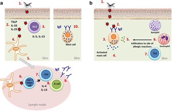
a.Sensitisation: (1) The Culicoides bites and injects saliva with proteins. (2) The cells of the epithelium react with production of TSLP, IL-33 and IL-25 alarmins, activating ILC2 cells. (3) ILC2 secrete IL-5 and IL-13. (4) – (6) Antigen presenting cell (APC) takes up the salivary gland protein (the allergen), migrates to the draining lymph node and under the influence of the proinflammatory cytokines presents the allergen to a CD4 T helper cell (Th). (7) – (9) The Th develops in to a Th2 cell producing IL-4 and IL-13 proinflammatory cytokines which instruct the B-cell to undergo Ig class switching to IgE. (10) IgE antibodies bind high affinity IgE receptors (FcerRI) on mast cells and thus sensitizing the horse.
b.Re-exposure: (1) - (2) When the horse is re-exposed to the allergens they bind to the mast cell-bound allergen specific IgE causing cross-linking between receptors, activating the mast cells to release inflammatory mediators causing inflammation and itching. (5) – (7). Mediators of the activated mast cells then recruit eosinophils, basophils and Th2 cells to escalate the reaction even further. From Jonsdottir et al., 2019.
Details of references in the text above.